


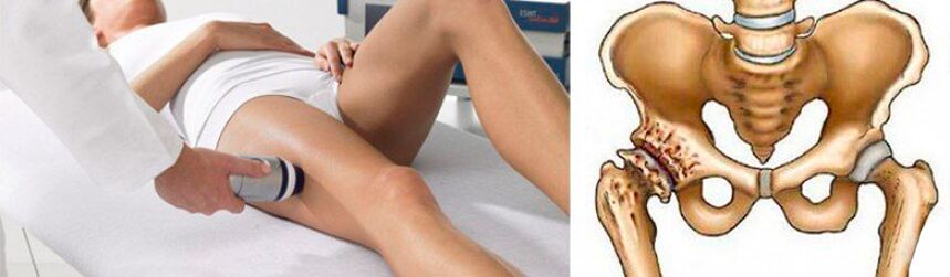
Coxarthrosis of the hip is a degenerative-dystrophic process occurring at the junction of the femoral head and pelvis. The disease is more typical in middle-aged and elderly people, although it can also occur in young people, including children. Usually, its development is preceded by trauma, as well as some inflammatory and non-inflammatory pathologies, and pain and stiffness with movement become the main sign of degenerative-dystrophic processes in the joints. Crotch. In the process of development, the disease goes through many stages, if in the early stages it can be treated conservatively, in the final stage, the treatment of hip osteoarthritis is only effective with surgery. Otherwise, the pathology will lead to serious disturbances or even complete immobility.
What is coxarthrosis of the hip joint and the mechanism of its development
Coxarthrosis, also known as osteoarthritis and joint deformity, is a complex disease of the hip joint (HJ), accompanied by the gradual destruction of cartilage. Over time, this leads to deformation of the surface of adjacent bones, as well as the formation of bony masses on them, known as osteoblasts.
According to statistics, musculoskeletal diseases account for about 12% of all diseases of the musculoskeletal system. In terms of frequency, it is second only to fibromyalgia, but the risk of disability is much higher.
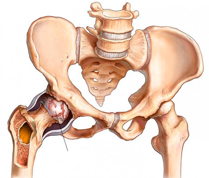
The hip joints are the two largest joints in the body. Each of them is formed by the femur and pelvis of the pelvis. The femoral head is located in the cup-shaped recess of the pelvis and moves freely in different directions. This structure of the joint makes it possible for you to be flexible and not flex, bend and flex, and also rotate the thigh.
To prevent uncomfortable movement, the surfaces of the bones that come in contact with each other are covered with an elastic layer called hyaline cartilage. It was he who allowed the femoral head to slide easily in the acetabulum. In addition, hyaline cartilage provides stability and cushioning to the hip joint during movement.
The entire joint is soaked in a type of shell known as a joint capsule. It contains a synovial membrane that synthesizes synovial fluid. It is she who lubricates the surface of the cartilage, ensures the flow of water and nutrients into it, i. e. is responsible for maintaining the normal structure of the cartilage tissue.
Above the joint capsule is a group of muscles of the femur and pelvis, with the help of which this joint is established for movement. The hip is also surrounded by a group of ligaments that ensure the stability of its position within physiological boundaries.
Because the hip joint is subjected to heavy loads on a daily basis, it is prone to rapid wear and tear. The risk of such changes greatly increases the influence of a number of adverse factors that are practically unavoidable in the modern world, but which are discussed below. This explains the high prevalence of coxarthrosis.
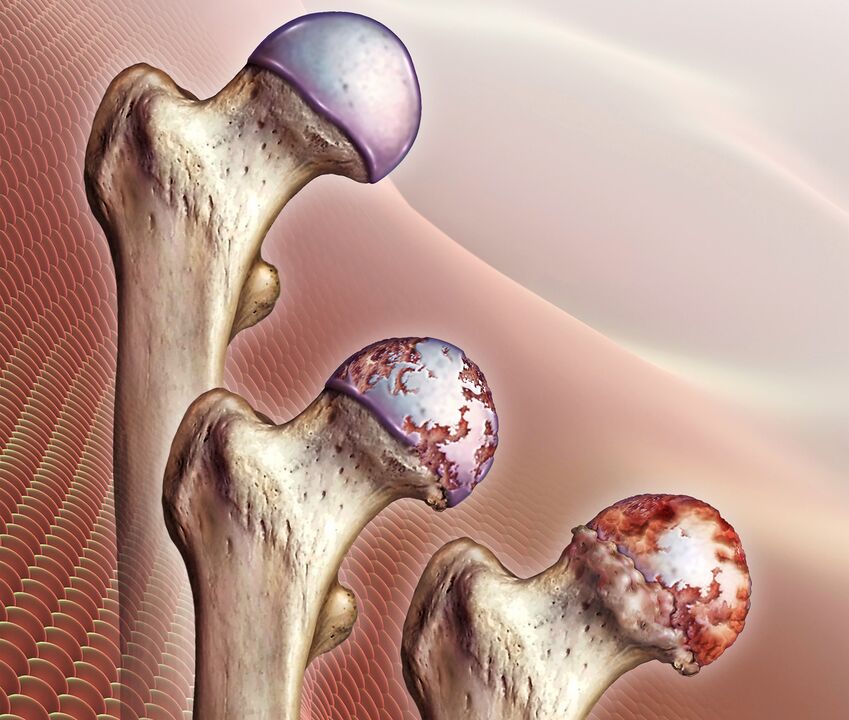
Due to the influence of negative factors, there is a violation of the production of synovial fluid. Gradually, its quantity decreases, and its quality composition also changes: it becomes viscous, dense and is no longer able to adequately nourish cartilage. This leads to acute nutritional deficiencies and gradual dehydration of hyalinized cartilage. As a result of such changes, the strength and elasticity of the cartilage tissue decreases, it peels, cracks and decreases in volume. All this prevents the smooth sliding of the femoral head in the pelvis, which leads to the appearance of signs of coxarthrosis in the hip.
Gradually, the space between the joints narrows, increased friction occurs between the bony surfaces, and the pressure of the bones on the hyaline cartilage increases. This leads to even greater injuries and abrasions, which cannot but affect the biomechanics of the hip joint and a person's health.
Damage to the hip joint not only negatively affects the biomechanics of the lower extremities, but also affects the entire motor apparatus. This often leads to disability.
As pathological changes progress, the hyaline layer gradually disappears completely, resulting in exposure of bony surfaces and increased load on the joints. During movement, the femoral head is no longer covered by anything and rubs directly against the surface of the pelvic pad. In addition to the fact that it severely limits mobility and causes unbearable pain, the bones press together, and at the same time flatten.
When bones are deformed, outgrowths of bone (osteogenesis) form on their surface. They can have sharp edges and seriously injure surrounding muscles. This leads to the appearance of severe pain in the groin, legs and buttocks. Therefore, the patient tries to free the affected hip and avoids movements in it unconsciously. The lack of adequate muscle load leads to progressive atrophy, exacerbating mobility problems. This leads to lameness.
Reason for development
Coxarthrosis of the hip can be primary or secondary. In the first case, it is not possible to find out the reason for its development, that is, the disease develops on its own for no apparent reason. Secondary coxarthrosis is the result of certain changes in the state of the musculoskeletal system or lifestyle features, namely:
- hip injuries, including fractures, dislocations, bruises, sprains or ruptures of surrounding ligaments, chronic minor injuries, etc. v. ;
- tiring physical work;
- sedentary lifestyle;
- fat;
- chronic infectious processes in the body;
- rheumatoid arthritis, gout, tendinitis, bursitis;
- endocrine diseases, metabolic and hormonal disorders, including diabetes mellitus;
- congenital malformations of the hip joint (dislocation, dysplasia);
- aseptic necrosis of the femoral head;
- pathologies of the spine of all kinds;
- genetic predisposition;
- tobacco addiction.
In the vast majority of cases, the development of coxarthrosis of the hip is due to inevitable age-related changes, and the presence of factors other than the above only increases the risk. appear and speed up progression.
Symptoms and severity
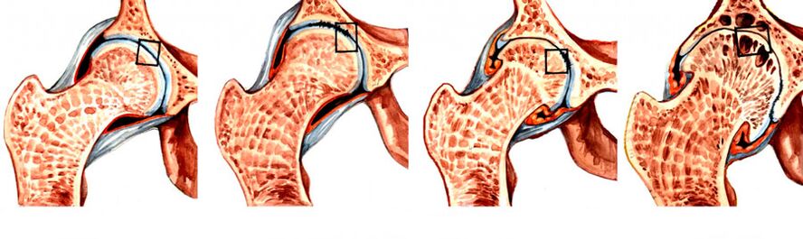
In the course of coxarthrosis, 4 levels of development are distinguished, of which 1 is the easiest. Initially, the disease may be asymptomatic or present with mild pain. They are more common after strenuous exercise, a long walk, or at the end of a busy day. At the first stage of the development of the disease, discomfort is often attributed to fatigue and is considered the norm. Therefore, it is extremely rare for coxarthrosis of the hip to be diagnosed at an early stage of development.
The recognizable signs of coxarthrosis begin to appear in the second stage of the progression, when the joint space is reduced by almost half, and the femoral head is displaced and deformed. In the third stage, the pain becomes unbearable and can disturb the patient even at night, they tend to spread to the hips, shins, groin and buttocks. Since there is practically no joint space and more osteoclasts are formed on the bone surface, independent movement in such situations is not possible. Therefore, the patient is forced to use a cane or crutches.
So, the main symptoms of coxarthrosis of the hip are:
- Limited mobility - initially, the patient may notice the appearance of difficulty performing rotational movements of the leg, but over time the stiffness and swelling of the HJ in the morning willjoin them. Because of them, a person needs several minutes to warm up and, so to speak, walk around to restore normal range of motion. Gradually, the patient becomes increasingly difficult to perform leg movements.
- A characteristic crunch - occurs when walking, as well as flexion or extension of the hip. It is a consequence of the friction of the bony surfaces against each other and with coxarthrosis accompanied by sharp or dull pain.
- Pain syndrome - pain that initially appears after exertion and subsides somewhat after a long period of rest. An acute attack can be provoked by weight lifting or hypothermia, as coxarthrosis is often complicated by the addition of synovitis. As the disease progresses, the pain becomes more frequent, lasts longer, and gets worse.
- Femoral spasm - is the result of compression of the nerves and weakening of the ligamentous apparatus, so the muscles contract compensatory to keep the femoral head in the femoral joint. In addition, muscle spasms may be provoked by additional bursitis.
- Lameness - occurs in the late stages of the development of the disease, since the deformation of the bone surface causes the appearance of spasticity of the flexor muscles. Therefore, a person cannot fully straighten the leg and hold it in this position. In addition, the patient may inadvertently limp to shift weight to the healthy side of the body, as this helps reduce pain intensity.
- Leg shortening - observed with grade 3 coxarthrosis. The lateral leg of the affected hip may be shortened by 1 cm or more due to narrowing of joint space, decreased muscle tone, and flattened femoral head.
In the final stage of development, the femoral head fuses with the acetabulum, resulting in complete immobility and disability.
Simultaneously, degenerative-dystrophic changes can be observed in one hip or both. Accordingly, characteristic symptoms will be observed on one side or both at the same time, but in the second case, their severity on the left and right side can be different.
Diagnose
The doctor may suspect the presence of coxarthrosis of the hip based on the patient's complaints, physical examination, and the results of functional tests. Be sure to measure the length of the leg when checking visually. For this, the patient is asked to stand up and straighten his or her legs as much as possible. The measurement is taken between the anterior axis of the pelvis and any bony structures of the knee, ankle, or heel. But if both hip joints are affected by coxarthrosis simultaneously, the data obtained will not be informative.
But since the typical symptoms of coxarthrosis can be accompanied by a number of other inflammatory and non-inflammatory diseases, instrumental examination is imperative for the patient to make an accurate diagnosis. It may be:
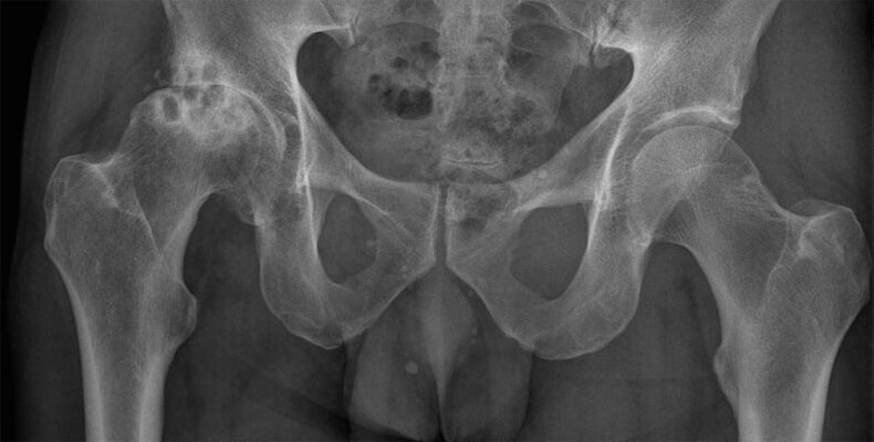
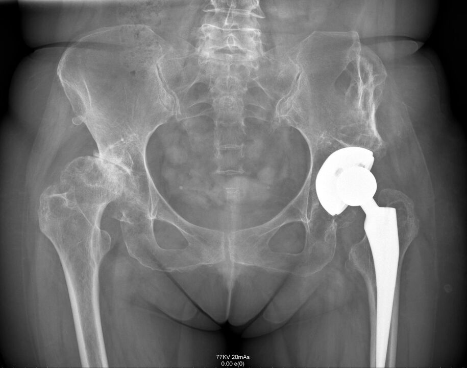
- CT or X-ray of the hip - the images show destructive changes in it, narrowing of the joint space, the formation of osteoblasts and deformation of the bone surface;
- MRI is the most informative examination method that allows you to accurately assess the nature of changes in the structure of cartilage, ligaments and the nature of blood circulation in the hip region.
Patients are also assigned laboratory tests to assess their general health and detect diseases that may cause coxarthrosis. It:
- UAC and OAM;
- blood chemistry;
- rheumatology test;
- Hip puncture by biochemical study.
The task of the diagnosis is to distinguish hip coxarthrosis from gonarthrosis (damage to the knee joint), as well as lens syndrome that occurs with osteonecrosis, as well as protrusion and herniated disc. In addition, the symptoms of coxarthrosis can resemble those of bursitis and an atypical episode of ankylosing spondylitis, requiring a complete examination to find the true cause of the pain. and limited mobility.
Conservative treatment
Conservative treatment of hip coxarthrosis is effective only in the early stages of the disease. It is selected for each individual patient and may include a whole range of different methods, each of which complements the others. Therefore, as part of treatment for coxarthrosis of the hip, patients may be prescribed:
- drug treatment;
- exercise therapy;
- physical therapy;
- plasmolifting.
For conservative treatment to be effective, it is necessary to eliminate the influence of several factors that contribute to the development of genital warts. If you are overweight, it is very important to lose as much as possible. This will reduce the load on the affected joint and the risk of progression of the degenerative-dystrophic process.
You should also give up smoking and normalize your exercise and sports regimen, avoid overload, but don't sit still all day. To prevent further destruction of the hip joint, you should wear special bandages and orthotics. They provide a secure fixation of the joint and support it during movement.
Medical treatment
The nature of drug therapy is strictly individualized. In most cases, the patient is prescribed:
- NSAIDs - drugs with simultaneous analgesic and anti-inflammatory effects (available in the form of tablets, injections and topicals);
- corticosteroids - drugs with a strong anti-inflammatory effect, prescribed if NSAIDs do not give a noticeable effect;
- chondroprotectors - contribute to the activation of the regeneration of cartilage tissue, but their effectiveness has not been proven;
- muscle relaxants - drugs that reduce muscle tone and eliminate spasms, necessary when spasms of certain muscles or muscle groups against the background of severe pain;
- preparations to improve blood circulation - most often used in the form of an injection solution and help to improve the nutritional properties of the tissues around the joints;
- B vitamins - have been shown to normalize the transmission of nerve impulses, which is especially important when nerves are compressed by deformed bone structures.
For acute pain that cannot be eliminated with the help of tablets, intra-articular or peri-articular blocks can be performed for the patient. They are performed exclusively by qualified medical personnel in a medical facility and consist of the introduction of anesthetic solutions into the joint cavity or directly around it.
exercise therapy
Exercise therapy is an effective method to address hypotonia and limited mobility. Thanks to a properly selected set of exercises, it is possible to increase the range of motion and reduce the severity of the pain. They also prevent muscle atrophy and help eliminate spasms if coxarthrosis is accompanied by compression of nerve fibers, which reflexively leads to spasm of individual muscles.
Therapeutic exercise classes can improve blood circulation in the area of the degenerative-dystrophic process. As a result, the nutritional quality of the diseased joint increases and the regeneration process accelerates.
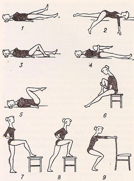
For each patient, a set of exercises should be developed individually by the therapist. At the same time, not only the degree of destruction of the hip joint is considered, but also the physical development level of the patient.
Physical therapy
Physical therapy and massage procedures have anti-inflammatory, analgesic, tonic, anti-edema effects. In addition, they help maintain normal leg muscle tone, preventing leg muscle atrophy.
With coxarthrosis of the hip, courses of 10-15 courses are prescribed:
- therapeutic ultrasound;
- acupuncture therapy;
- laser therapy;
- electrophoresis;
- super optoelectronics;
- UHF;
- paraffin treatment.
In addition, many patients are offered mud therapy. Such procedures have a positive effect only at the first stage of the development of coxarthrosis of the hip joint or during rehabilitation after surgical treatment. Thanks to the therapeutic mud, it is possible to improve the quality of blood circulation and accelerate the recovery of mobility of the affected joint.
Plasmolifting
Plasmolifting or PRP-therapy is a procedure that involves the introduction of platelet-rich plasma from the patient's own blood into the hip joint space. This allows you to activate the hyaline cartilage repair process.
However, according to some scientists, such a procedure can cause the formation of malignant tumors. This view is based on the fact that plasmolifting promotes the formation of a large number of stem cells, the effect of which on the body has not yet been fully studied.
Surgical treatment of coxarthrosis of the hip joint
Despite significant hip discomfort, many people seek medical help too late, when pathological changes in the joint reach 3 or even 4 severity and function is impaired. unrecoverable.
With advanced disease, surgery is necessary. Only timely surgical intervention will restore normal mobility and save the patient from severe pain, that is, the quality of life of the person is significantly improved. There are no drugs or physical therapy procedures that can restore severely damaged cartilage. At best, pain medications and intra-articular injections can relieve pain. But this will be a temporary phenomenon, after which the pain will recur with equal or even greater strength.
Indications for hip surgery are:
- the disappearance of internal space;
- persistent pain in the hip joint, which cannot be relieved;
- severe movement disorders;
- hip fracture.
Depending on the severity of the joint destruction and bone deformity, patients may be offered different forms of surgical treatment, namely:
- arthrodesis;
- endospores;
- osteopathy.
Arthrodesis
Joint grafting is an affordable surgery that involves the secure fixation of bones and joints with metal plates. As a result, the joint is completely immobilized. So, with the help of arthrodesis, it is only possible to correct the supporting function of the legs, eliminate pain, let alone restore mobility or significantly improve quality of life.
Today, arthrodesis is practically unused, since it deprives a person of the opportunity to completely move.
Endoscopic
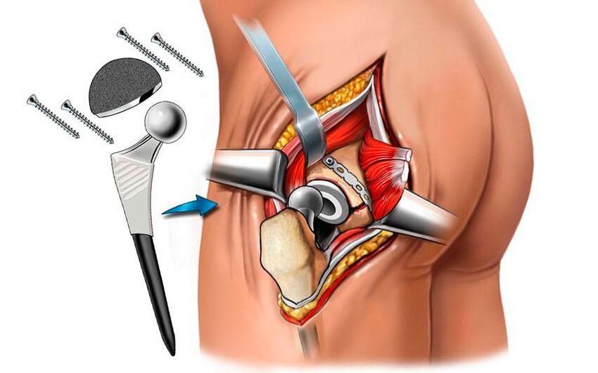
Arthroscopy with arthroplasty is the only way to completely solve the problem of hip osteoarthritis by restoring all function and mobility of the hip. This is a high-tech approach to solving the problem of coxarthrosis, allowing you to completely forget about it within 15–30 years, as well as pain and mobility limitations. Thanks to the use of modern endoscopy, it is possible to completely restore the motor support functions and bring the patient a normal life.
The surgery involved removing the femoral head and part of its neck. Surgical preparation for the acetabular bed is also performed, which includes the removal of osteocytes, its surface alignment, and excision of the necrotic tissue. Endogenous drugs may even be used to treat elderly patients with hip coxarthrosis.
The surgery is done under general anesthesia and takes about an hour. Depending on the severity of the degenerative-dystrophic process, surgery can be performed using one of the following methods:
- superficial - involves grinding the acetabulum and femoral head with an additional coating of smooth implants that replace the destroyed hyaline cartilage (this method is rarely used due to the possibility of inflammation in the periarticular tissues);
- unipolar - removal of the femoral head and its replacement with an organ (used when cartilage is preserved on the surface of the acetabulum and only the femoral head is destroyed);
- bipolar - similar to the previous technique, differing only in the design of the medical device used, which has a lower coefficient of friction and provides smoother motion in the joint bed;
- Total is the most effective and safest method to solve the problem of hip osteoarthritis, which involves complete removal of the femoral head with partial capture of its neck, as well as the tarsal bones, and their replacement. by a complete artificial. coupling.
As a result, the patient may be recommended for a variety of endotracheal intubations. Most hip replacement devices are made in the US and UK. For their manufacture, chemically and biologically inert metals are used: alloys of cobalt, chromium, titanium. Often ceramics are also used. In most modern models, a polymeric spacer is additionally used, which provides natural absorption, stability and sliding properties to the artificial TBS.
When performing medical cosmetic surgery, the success rate of surgery is almost 100%.
After surgery, antibiotics are prescribed to prevent the development of infectious complications, and the stitches are removed after 10 days. The postoperative scar was approximately 8 cm in size, and the patient was discharged home. Rehabilitation after endoscopy is simple, but still requires physical therapy, massage and exercise.
cut bone
Osteotomy is a surgical intervention that is a temporary measure before arthroscopic hip replacement. The essence of the operation is to align the axis of the femur due to intentional fracture. The resulting debris is placed in the most appropriate position, thus slightly lifting the diseased joint. As a result, it is possible to temporarily reduce pain severity and improve mobility.
As such, coxarthrosis of the hip is a rather formidable disease that can completely deprive a person of the opportunity to move independently. It progresses over a long period of time, and its symptoms, especially in the early stages, are often considered normal by the patient after exertion. But it is this that speaks to the insidiousness of this disease, because only at an early stage of development can it be treated in a non-surgical way. But if the degenerative-dystrophic process has completely destroyed the hyalinized cartilage and leads to the exposure of the bone surfaces, and even their flattening, then only surgery can help the patient. Fortunately, the modern level of medicine and surgery, in particular, can help to fully restore the normal state of the hip and its functions.